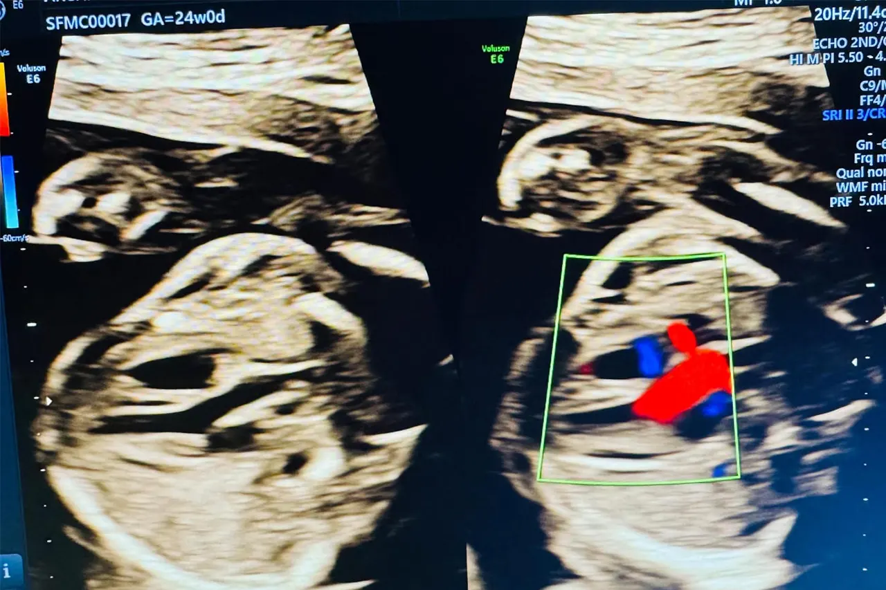
Fetal Echocardiography
A detailed ultrasound examination of your baby’s heart to assess structure, rhythm, and blood flow—helping detect heart conditions early during pregnancy.

What Is Fetal Echocardiography?
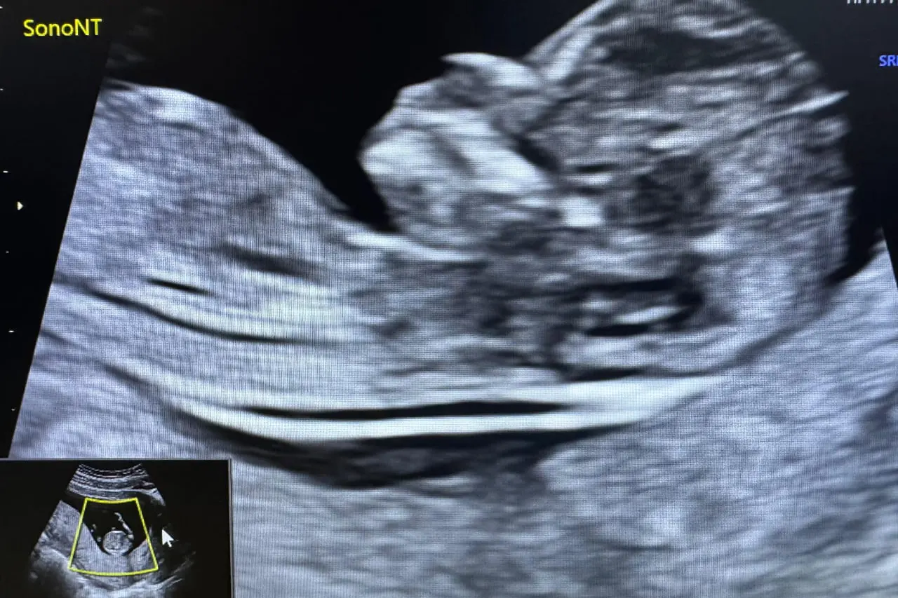
Fetal echocardiography is a specialized ultrasound scan used to closely examine the structure and functioning of the unborn baby’s heart. This scan provides detailed images of the heart chambers, valves, blood vessels, and heart rhythm, allowing early detection of congenital heart abnormalities.
It is a safe, non‑invasive procedure typically performed during the second trimester and may be recommended based on medical history, screening results, or specific risk factors.
Why Is Fetal Echocardiography Done?
- Detect congenital heart defects before birth
- Evaluate heart structure, blood flow, and rhythm
- Confirm findings from routine ultrasound scans
- Assess babies with suspected cardiac abnormalities
- Enable early planning and appropriate medical care
When Is Fetal Echocardiography Recommended
- Abnormal findings on routine anomaly or NT scan
- Family history of congenital heart disease
- Maternal conditions such as diabetes or infections
- Exposure to certain medications during pregnancy
- Multiple pregnancies or IVF conception

What the Scan Evaluates?
- Heart chambers and wall structure
- Heart valves and major blood vessels
- Blood flow patterns within the heart
- Heart rhythm and function
- Signs of congenital cardiac abnormalities
Opening Hours
Mon – Sat: 10.00 AM – 4.00 PM
Sun: 09.00 AM – 4.00 PM
Friday: Closed
Emergency: 24 hours
Contact Us
Our Clinic Gallery
Explore our facility and see where we provide exceptional care